
AVM Awareness Month: What is an AVM, and why does it matter?
October is AVM Awareness Month, a chance to talk about a condition most people have never heard of until it affects someone they love.
An AVM, or arteriovenous malformation, is a tangle of blood vessels that hasn't formed the way it should. Many people live with one for years without knowing. For others, the first sign is a bleed on the brain.
At the Natalie Kate Moss Trust, brain haemorrhage is why we exist. AVMs are one of the causes, and they're often the reason a brain haemorrhage happens to a child or young adult. So this month, we want to help more people understand what an AVM is, what to look out for and where to turn for support.
What is AVM Awareness Month?
AVM Awareness Month takes place every October. It's best established in the United States, where October is formally recognised each year as a month to raise awareness of arteriovenous malformations. Awareness shouldn't stop at a border, so we're marking it here in the UK too.
The aim is simple: help more people recognise the signs, support those living with an AVM and back the research that could improve treatment.
What is an AVM?
AVM stands for arteriovenous malformation.
It's a tangle of abnormal blood vessels that connects arteries directly to veins.
Normally, arteries carry oxygen-rich blood away from the heart at high pressure. That blood passes through a network of tiny vessels called capillaries, which slow it down and deliver oxygen to the tissue around them. Veins then carry the blood back to the heart at a much lower pressure.
In an AVM, the capillaries are missing. Blood rushes straight from the arteries into the veins through the tangle. That causes two problems. The tissue around the AVM can miss out on the oxygen it needs. And the vessels inside the tangle are under more pressure than they were built for, which makes them weaker and more likely to bleed.

AVMs can form anywhere in the body, but they're most often found in the brain and spinal cord.
One in the brain is called a brain AVM or cerebral AVM, and that's the type we're focusing on here.
A few things worth knowing:
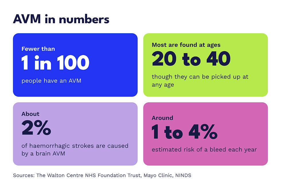
AVMs are rare. They affect fewer than 1 in 100 people.
They aren't cancer. An AVM isn't infectious and doesn't spread to other parts of the body.
Most are found between the ages of 20 and 40, although they can be picked up at any age.
No two are the same. Some are tiny, others cover a larger area of the brain.
(Source: The Walton Centre NHS Foundation Trust)
What causes an AVM?
The short answer is that nobody knows for sure. Most AVMs are thought to be present from birth, forming as the blood vessels develop, though some may appear later in life. (Source: National Institute of Neurological Disorders and Stroke)
An AVM isn't caused by anything you did or didn't do.
AVMs don't usually run in families.
In rare cases they're linked to an inherited condition called hereditary haemorrhagic telangiectasia (HHT).
You can read more about family history in our blog, Are brain haemorrhages hereditary?
AVM symptoms: what to look out for
Some people with a brain AVM have no symptoms at all.
In around 15% of cases, the AVM is found by chance during a scan for something else.
When symptoms do appear, they depend on where in the brain the AVM sits.
They can include:
Seizures
Headaches, which can feel like a migraine or keep returning in the same spot
Weakness, numbness or tingling in one part of the body
Problems with your vision
Difficulty speaking or understanding words
Dizziness or trouble with balance and coordination
A whooshing or pulsing sound in your head, in time with your heartbeat
Memory problems or confusion

All of these have other causes too, and most are far more common than an AVM. But if something is new, keeps coming back or just doesn't feel right, speak to your GP.
When an AVM bleeds
The biggest risk with a brain AVM is that it ruptures and bleeds into or around the brain. This is a brain haemorrhage, also known as a haemorrhagic stroke.
More than half of brain AVMs are only discovered after they've bled.
Around 1 in 5 are found because of seizures.

For an AVM that hasn't bled, estimates of the risk range from around 1% to 4% each year, depending on the study and on the AVM itself.
After a bleed, the risk is higher for the next couple of years. (Sources: NINDS, The Walton Centre)
Brain AVMs account for about 2% of all haemorrhagic strokes. That may sound small, but in children and young adults who have a brain haemorrhage, an AVM is often the cause. (Source: Mayo Clinic)
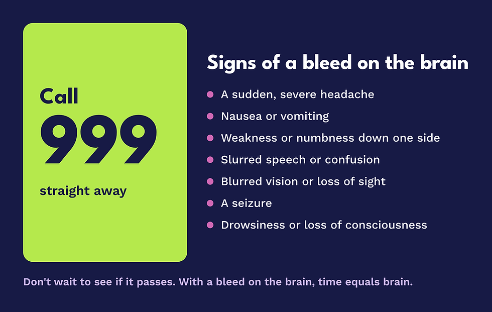
Call 999 straight away if you or someone with you has:
A sudden, severe headache, often called a thunderclap headache
Nausea or vomiting
Weakness or numbness, especially down one side of the body
Slurred speech or confusion
Blurred vision or loss of sight
A seizure
Drowsiness or loss of consciousness
Don't wait to see if it passes. With a bleed on the brain, time equals brain. Read more about the warning signs and symptoms of a brain haemorrhage.
How is an AVM diagnosed?
An AVM is diagnosed with brain scans. You may have one or more of these:
CT scan. Often the first scan in an emergency, as it shows bleeding in the brain quickly.
MRI scan. Gives a detailed picture of where the AVM sits and what's around it.
Angiogram. A thin tube is passed through an artery, usually from the groin or wrist, and dye is injected so doctors can see the exact shape, size and blood supply of the AVM on X-ray.
How is an AVM treated?
Every AVM is different, so treatment is planned one person at a time.
In the UK, a team of specialists meets to review your scans and agree the safest plan, then talks it through with you. Nothing is decided without you.
The main options are:
Monitoring
If the risks of treatment outweigh the benefits, your team may recommend regular scans instead.
Surgery
A neurosurgeon removes the AVM through an opening in the skull. This tends to suit smaller AVMs nearer the surface of the brain.
Embolisation
A thin tube is guided through the blood vessels to the AVM, and a glue-like substance is used to block it off from the inside. It's often used alongside surgery or radiosurgery.
Stereotactic radiosurgery
Highly focused beams of radiation, such as Gamma Knife, are aimed at the AVM so the vessels gradually close over months or years. No incision is needed.
Many people have a combination of these. Some AVMs sit in parts of the brain where treatment would be too risky, and are monitored instead.

Can an AVM be prevented?
You can't prevent an AVM from forming.
What you can do is look after the blood vessels you have.
High blood pressure puts extra strain on the blood vessels in your brain, and vessels that are already weakened are more vulnerable to it. It's one of the leading causes of brain haemorrhage overall, and it's the one we can all do something about. That's why we spend so much time encouraging people to know their numbers.
If you've been diagnosed with an AVM, your specialist team is the best source of advice on what's right for you, including blood pressure, exercise and pregnancy.
How to get involved this AVM Awareness Month
Share this article. One share could be the reason someone recognises the signs.
Learn the warning signs of a brain haemorrhage, and what to do.
Get your blood pressure checked. It takes a few minutes at a pharmacy, your GP surgery or one of our awareness days.
Fund research. £33 funds one hour of brain haemorrhage research.
Listen and learn. Survivors share their stories on our Prevent The Preventable podcast.
Help us change what happens next
At the Natalie Kate Moss Trust, we work to prevent brain haemorrhages and fund research at The University of Manchester to develop life-saving treatment for when they do happen.
This AVM Awareness Month, please donate what you can. Every hour of research brings better treatment closer.
This article is for general information and isn't a substitute for medical advice. If you're worried about your health, speak to your GP or call NHS 111. In an emergency, always call 999.
FAQs
When is AVM Awareness Month?
AVM Awareness Month is in October every year.
What does AVM stand for?
AVM stands for arteriovenous malformation. "Arterio" refers to arteries, "venous" to veins, and "malformation" means the vessels haven't formed in the usual way.
What is an AVM in the brain?
A brain AVM, also called a cerebral AVM, is a tangle of abnormal blood vessels in the brain that links arteries straight to veins. Because the blood skips the tiny capillaries that would normally slow it down, the vessels are under extra pressure and can bleed.A brain AVM, also called a cerebral AVM, is a tangle of abnormal blood vessels in the brain that links arteries straight to veins. Because the blood skips the tiny capillaries that would normally slow it down, the vessels are under extra pressure and can bleed.
Are AVMs rare?
Yes. AVMs affect fewer than 1 in 100 people, and many of those people never know they have one.
Are AVMs dangerous?
They can be. Many people with an AVM have few symptoms or none. The main danger is a bleed on the brain, which is a medical emergency. That's why an AVM is always assessed by a specialist team once it's found.
Is an AVM hereditary?
Usually not. Most AVMs happen by chance. In rare cases they're linked to an inherited condition called hereditary haemorrhagic telangiectasia (HHT).
What is an AVM rupture?
An AVM rupture is when one of the weakened vessels in the tangle bursts and bleeds into or around the brain. It causes a brain haemorrhage, also called a haemorrhagic stroke, and needs emergency treatment. Call 999.
Is an AVM the same as a brain aneurysm?
No. An aneurysm is a bulge in a weak spot on the wall of a single blood vessel. An AVM is a tangle of vessels connecting arteries and veins. Both can cause a brain haemorrhage, and some people with an AVM also develop aneurysms.
Can an AVM be treated?
Often, yes. Treatment can involve surgery, embolisation, stereotactic radiosurgery or a mix of these. Where treatment would carry too much risk, the AVM is monitored with regular scans instead.
What is the life expectancy of someone with an AVM?
There isn't a single figure. It depends on the size and position of the AVM, whether it has bled and how it's managed. Many people with an AVM have few or no serious problems from it. Your specialist team can talk you through what your own scans show.

